
TSU biophysicists and colleagues from India and Taiwan have developed new approaches for studying the tissues of patients with serious diseases, such as diabetes mellitus, and oncopathology. The scientists used nonlinear optical microscopy, which provides a large amount of new data on changes in the patients’ tissues. This data will help create effective treatments and identify, for example, the causes of poor wound healing. The research is presented in the Journal of Applied Physics (Q2), published by the American Institute of Physics since 1931. The article was chosen as the central theme of the issue and placed on its cover.
- There are two points important in the diagnosis of diseases, based on the analysis of biological tissues: information about the structure and molecular composition, which means the composition and quantity of proteins and other substances, - explains Yury Kistenyov, one of the authors of the article, head of the TSU Laboratory of Biophotonics, executive director of the TSU Institute of Biomedicine. - Traditional microscopy in most cases gives information only about the structure of tissues. But the advent of powerful femtosecond lasers has led to the emergence of nonlinear microscopy methods that enable studying subtle effects in biological matter.
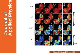
Using these methods, scientists can see the distribution of various proteins (elastin, collagen, and others), evaluate the metabolism within the cell (for example, energy metabolism), and examine other parameters. In the article, the authors describe the wide diagnostic capabilities that open up the use of individual methods of nonlinear microscopy and their combination.
As one example, TSU scientists cite the results of studies related to wound healing in patients with lymphedema, a severe pathology that often develops after the radical treatment of some oncological diseases.
- When it comes to healthy tissues, there are no problems with wound healing. But in the presence of severe pathologies, for example, diabetes mellitus, regeneration has its characteristics and is often delayed and proceeds with complications, says Yury Kistenyov. - To effectively solve such problems, we need to know what deviations from the norm develop in the tissues of such patients. They can be detected by nonlinear microscopy methods.
Thus, in the case of lymphedema in an experiment on rats, TSU biophysicists revealed disarrangement of collagen distribution and other violations of the molecular composition, which could be the cause of poor wound healing.
The results presented in the article, “Label-free Multimodal Nonlinear Optical Microscopy for Biomedical Applications”, will help other scientists in medical physics create new diagnostic technologies. The data obtained using biophotonic methods will be the basis for new therapeutic approaches.
The importance of the research work of the group of scientists from Russia, India, and Taiwan was also noted by the editors of the Journal of Applied Physics in, choosing the article for the cover of the issue.