
TSU neuroscientists, using a new noinvasive method, were the first in the world to trace the death and recovery of nerve cells and axons after a brain stroke. The data were obtained in a rat model of ischemic stroke. Using a new approach to MRT (magnetic resonance tomography), the researchers tracked changes in the brains of animals that survived a vascular accident for three months. They found that the new axons were trying to restore the lost connection between nerve cells.
The diagnostic technology created in the TSU Laboratory of Neurobiology under the supervision of Vasily Yarnykh, its scientific advisor, professor at TSU and the University of Washington, became a tool for research. Using special procedures for the mathematical processing of MRT data, the scientists obtained myelin maps - images of the brain, reflecting the number of sheaths of nerve fibers in the same way as the terrain is depicted on geographical maps. Myelin maps reveal microscopic changes in brain matter that cannot be detected with conventional MRT.
- Two experiments were carried out: in the first, the state of the animals was monitored for ten days after the stroke, - says Marina Khodanovich, head of the TSU Laboratory of Neurobiology. - In the second, multiple scans of the rodent brain were carried out over three months. In both cases, we looked at what happens to neurons and axons (processes of nerve cells), along which impulses go from one nerve cell to another.
At the same time, the scientists carried out a quantitative assessment of myelin - the main substance in the composition of the nerve sheaths. The correct transfer of information between the cells of the nervous system depends on its state. Studies have shown that after a stroke in the area of the brain where there was a violation of the blood supply, neurons and axons die, but some of the axons remain alive, albeit demyelinated. It also turned out that this process is reversible - over time, myelin is restored in the nerve sheaths.
There is also another phenomenon: from healthy neurons from intact areas, in which axons have died and therefore communication with other nerve cells is broken, new axons grow. They can pass through the ischemic region, reaching healthy neurons and forming new neural networks. If a new connection is formed, and nerve impulses begin to pass along the axon, this is a signal for the formation of a new myelin sheath. The germination of new nerve endings in rats occurs rather quickly, after two to three months.
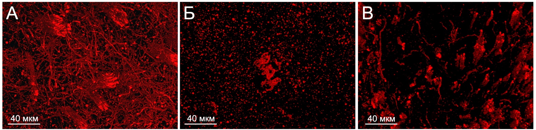
A - normal myelin sheaths of axons in the rat brain
B - the destruction of the myelin sheaths in the ischemic focus 10 days after the stroke
C - the restoration of the myelin sheaths of axons 30 days after the stroke
- Apparently, the new conducting network does not fully reproduce the one that was there before the damage, but, nevertheless, the axons are trying to establish the lost connection between the cells, - says Marina Khodanovich. - Our next task is to find out how the restoration of the network affects the restoration of various functions: perception, memory, fine motor skills, and others. To do this, it is necessary to understand how the structure of the nervous network has changed, and compare this with the restoration of functions. This will be the focus of our further research.
The scientists intend to use a combination of two methods of magnetic resonance tomography: MPF mapping and tractography, which help to assess the pathways of the nervous system. This will help to see from the inside the processes of brain recovery after a stroke and compare them with the dynamics of rehabilitation of sensory, motor, and cognitive abilities. The new approach is promising both for monitoring the condition and predicting the dynamics and volume of restoration of lost functions.
Along with this, the neuroscientists plan to obtain new fundamental data by studying the molecular mechanisms of neuron and axon repair. Scientists will try to answer many questions for the first time, in particular, to determine how a healthy axon finds its way to the cell with which it needs to communicate, and whether it always manages to do it. Researchers expect to find targets, impact on which will stimulate recovery processes and accelerate the return of patients to a normal quality of life.